Better screening could reduce the use of invasive heart tests, based on a recent study that used non-invasive CT imaging as a gatekeeper for selecting patients at high risk for heart disease. Such imaging could help reduce use of invasive coronary angiographies in patients with healthy arteries, saving both time, money and added risks for patients.
Published in the Journal of the American College of Cardiology: Cardiovascular Imaging, this study was conducted at 22 centers in North American, East Asia, Europe and India. It included more than 1,600 patients with suspected heart disease, all of who required further testing to assess the heart’s arteries.
Through the trial, roughly half of patients received standard care, which includes invasive coronary angiography to check for narrowing or blocked arteries. During a coronary angiogram, a colored die is injected into the heart’s blood vessels and X-ray imaging is used to identify blocked arteries. The test is generally safe and is used to diagnose (and potentially treat) heart disease. However, angiography carries small risks of serious complications and often yields normal results for most patients.
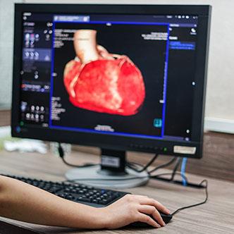
For this reason, half of study participants underwent a “pre-screening” prior to angiography. This screening included a non-invasive imaging test called computed tomography angiography (CTA), which uses CT imaging to assess the heart’s arteries. The CTA was used to narrow down which patients were most likely to have heart disease and in need of the invasive angiogram.
After these tests, researchers followed participants for one year to track outcomes like heart events, hospitalizations, emergency heart procedures and death.
The average age of participants was 60 and nearly half were women.
Overall, only 23% of participants in the screening group went on to have coronary angiograms—25% of whom had normal test results. A total of 13% of participants required revascularization procedures to clear any blockages in their arteries.
All participants in the comparison group underwent coronary angiograms, 61% of whom had normal results and 18% of whom required revascularization procedures.
Experts note that after following participants for one year after their testing, risk of heart events was the same in both groups.
It’s important to note that this study only included stable patients with suspected heart disease, who did not require immediate testing and treatment. To qualify for the study, participants had symptoms of heart disease or an abnormal stress test but were not being evaluated for heart attack or emergency conditions. Therefore, findings don’t apply to patients with more unstable symptoms or conditions.
What findings show, according to authors, is that CT imaging helps reduce invasive testing without compromising safety in patients with suspected heart disease. In this study, the additional imaging helped cut unnecessary angiograms in half and reduced the number of patients requiring revascularization. As authors highlight, these benefits were achieved without affecting participants’ risk for heart events after one year.
Additional research is needed to confirm the long-term effects of this screening strategy. However, findings are encouraging when it comes to helping reduce unnecessary procedures and risks in patients with suspected heart disease.