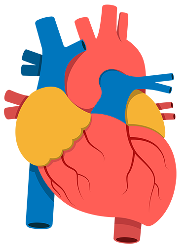
Your Heart Is Special
Learning About Your Heart Can Help
Find ways to manage and live well with your condition.
Deciding What's Right for You
If you have a heart condition, you may need to make decisions about tests, procedures or medications. The American College of Cardiology has tools to help you talk with your care team, understand your options, and choose what’s right for you.

Share Materials With Your Patients Search All Tools
To support health care conversations between patients and their clinicians, the American College of Cardiology created tools to strengthen their partnership – not just on specific decisions but over a lifetime.
Start Exploring Some Today